
Example Procedure
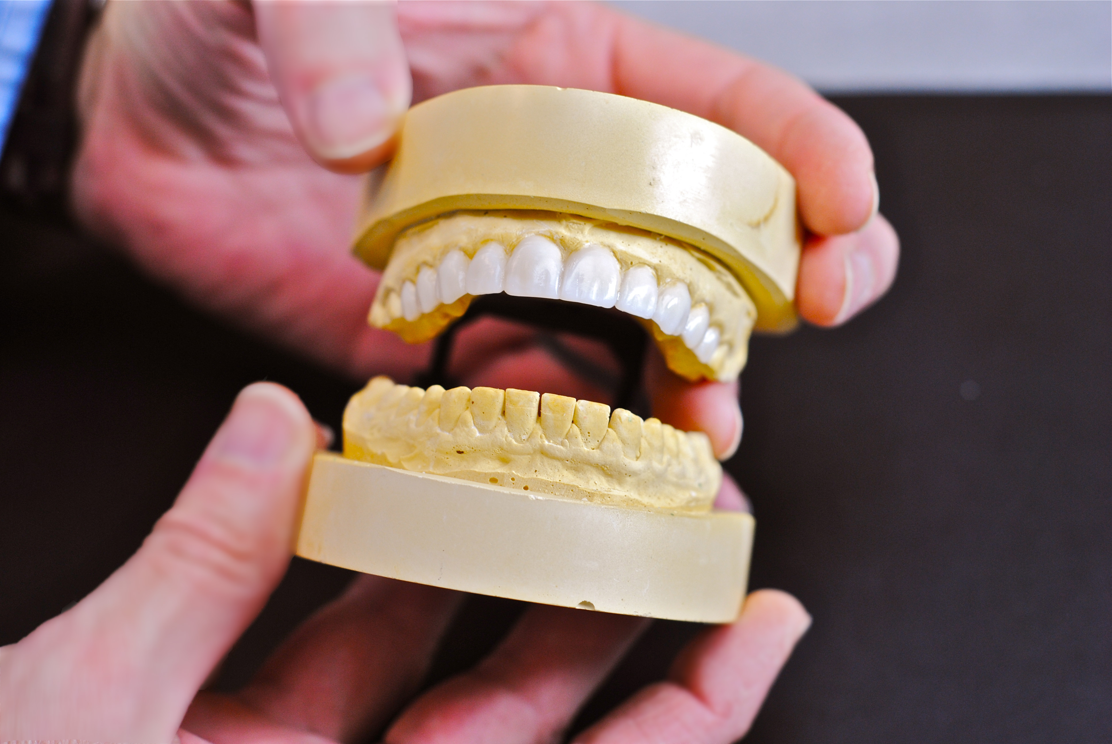
The goal of a Diagnostic wax-up is to pre-determine the approach to the restoration. Once this has been determined, we do the wax-up by adding wax and or removing from the tooth structure (stone model). Reductions to the stone model will be marked in red, or a stent can be made to indicate the reductions made on the model.
After the wax-up is completed, it will be duplicated and poured in stone to fabricate a stent which will be used to make the provisional s in the mouth. Once the patient has approved all the changes, we can move to the next step. First an impression is taken of the approved
Provisionals and a new Face bow registration is done. The new model of the provisional can be mounted in the Dental office, or the Face bow and the model can be sent to us.
Procedure Pictures
Coming Soon!
Pictures featuring Diagnostic Wax-ups are coming soon.
Requirements
- Full mouth impression including the vestibule
- Centric relation bite (CR)
- Face bow or Face bow mounted upper model
- Clinical finding of individual teeth and which teeth will have to be extracted
- Diagnostic photos;
- Full face smile (This photo should have only the full face and shoulder in it)
- Close-up smile (Should be from nose to chin)
- Close-up at rest (lips slightly apart – from nose to chin)
- Close-up high smile (This will show the relationship between the upper lip and the gingival contour)
- Close-up profile – patient smiling (This will show the relation between the upper incisal edge and the lower lip)
- Close-up, top to down, nose to chin – patient smiling (This will show the upper arch against the lower lip)